Collotheca rotifer unveils its feeding web
Collotheca is a genus of rotifer that feeds by extending long hair like structures called setae, in which it traps its prey. In the case of this sessile (that is, not swimming) species ornata, the prey is largely made up of bacteria and tiny algae.
Here you can see the full process of opening up its mouth, made up of 5 total lobes (not all seen clearly in this focal plane), and extending all its setae, before being startled and contracting back into its hiding spot. Even the lightest touch on the microscope stage would cause it to contract; those are some sensitive whiskers.
Technical details
Olympus BH2, SPlan 40x 0.70, 1.25x intermediate lens, 2.5x NFK, Canon EOS 5D Mark II. Condenser stopped down significantly to add contrast to the setae. Pond water sample from park in Paris 16eme.
Tiny Cyclidium scooping up its dinner
Had a chance to take a long and chill look at this tiny Cyclidium sp. demonstrating its unique way of feeding (at least compared to other ciliates I normally observe).
As it spins, you can just barely see a large, transparent sheet-like membrane protruding from along its side. This is the “paroral membrane” that acts like a scoop, sweeping food directly into its mouth.
Technical details
Olympus BH2, SPlan 100x 1.25 oil, 1.25x intermediate lens, NFK 2.5x photo eyepiece, Canon EOS 5D Mark II. Pond water sample from a park in Paris 16ème.
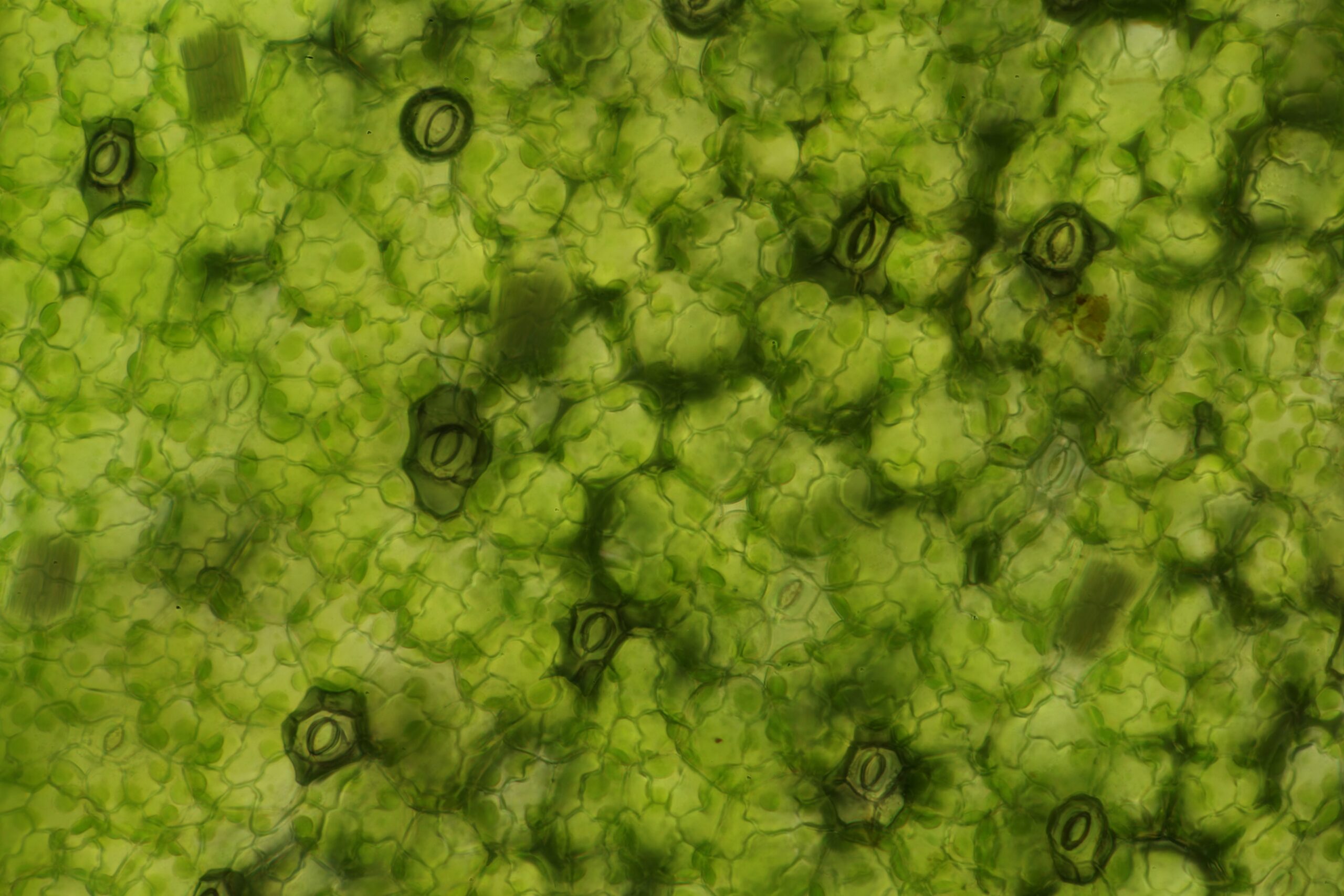
The microscopic lungs of duckweed
On the top of every frond of duckweed you can find these little mouth shaped holes called stomata; these allow this aquatic plant to breathe while keeping the vast majority of itself underwater. Duckweed is a fantastic place to look for all sorts of microscopic organisms, many of which anchor themselves to the roots.
Technical details
Olympus BH2, SPlan 40x 0.70, NFK 2.5x photo eyepiece, Canon EOS 5D Mark II. Focus stack of 10 individual images prepared using Picolay. Pond water sample from park in Paris 16eme.
Monstrously large Euglenid
Euglenids are some of my favourite creatures. Most of them swim along with the help of a flagella that they whip around in front like a propellor.
But this one can barely get off the surface. This absolutely massive Euglenid appears to be roughly 200 µm, much greater than the types I normally see. Based on its very short flagellum and worm-like shape and behaviour, I’m tentatively identifying it as Euglena ehrenbergii. Shown here in timelapse to emphasize the twisting motion and shape morphing, known as metaboly.
Also visible here are Coleps, Phacus, Closterium, diatoms, some small loricate euglenids, and a very quick drive by of some species of rotifer.
Technical details
Olympus BH2, SPlan 10x 0.30, Olympus 1.25x analyzer, NFK 2.5x photo eyepiece, Canon EOS 5D Mark II. Video is sped up 4x with 1.5 digital zoom, edited with OpenShot. Sample from pond in park, Paris 16eme.
Lovely, leaf-shaped Phacus drifting along
Phacus is a beautiful genus of Euglenid, leaf-shaped with a prominent eye spot. This species longicauda is characterized by a long spine and a markedly curved or twisted body shape, which you can see clearly when it rotates along its long axis (challenging to keep in focus however).
The flagellum should be about the length of the body but I had difficulty resolving it at any magnification. It’s just barely visible protruding from the lip at a few points in the clip.
Technical details
Olympus BH2. SPlan 40x 0.70, SPlan 100x 1.25 oil, 1.25x intermediate lens, 2.5x NFK photo eyepiece. Canon EOS 5D Mark II. Sample from pond in park, Paris 16eme.
Hairy belly gets itself stuck
Gastrotrichs come by their name honestly and are commonly known as hairy bellies (derived from Ancient Greek γαστήρ/gastēr for stomach and θρίξ/thrix for hair).
This is an example of Chaetonotus, a genus of gastrotrich, that seemed to get itself stuck on a piece of debris (like a poor kid putting his head through a fence). This gave me a chance to oil the slide and take a closer look at its scales and hairs. The pharynx and digestive system are also clearly visible.
Technical details
Olympus BH2, SPlan 40x 0.70 & SPlan 100x oil 1.25, 1.25x intermediate lens, 2.5x NFK photo eyepiece. Canon EOS 5D Mark II. Pond water sample from park in Paris 16eme.
Amoebas require patience
When searching for creatures it’s normally movement that catches my eye, but that doesn’t work for amoeba. They take life very slow.
This timelapse compresses about a 5 minute journey across the slide to just about 30 seconds.
Technical details
Olympus BH2, SPlan 40x 0.70, 1.25x intermediate lens, 2.5x NFK photo eyepiece, Canon EOS 5D Mark II. Video edited with OpenShot (brightness, contrast, desaturation, 8x speed for timelapse). Pond water sample from park in Paris 16eme.
Dozens of Vorticella take up residence on a duckweed root
Vorticella is a genus of single celled, bell-shaped ciliates that spend most of their lives filter feeding, anchored by a tether that snaps back like the world’s tiniest spring. The snap happens so fast it’s almost impossible to see – at a human scale, it would mean going from a standstill to the speed of sound in one second.
While Vorticella are solitary organisms, they tend to cluster together, as seen here in a long slow pan along the root of a single piece of duckweed.
Technical details
Olympus BH2, SPlan 10x 0.30, 1.25x intermediate lens, 2.5x NFK photo eyepiece, Canon EOS 5D Mark II. Sample from pond in park, Paris 16eme.
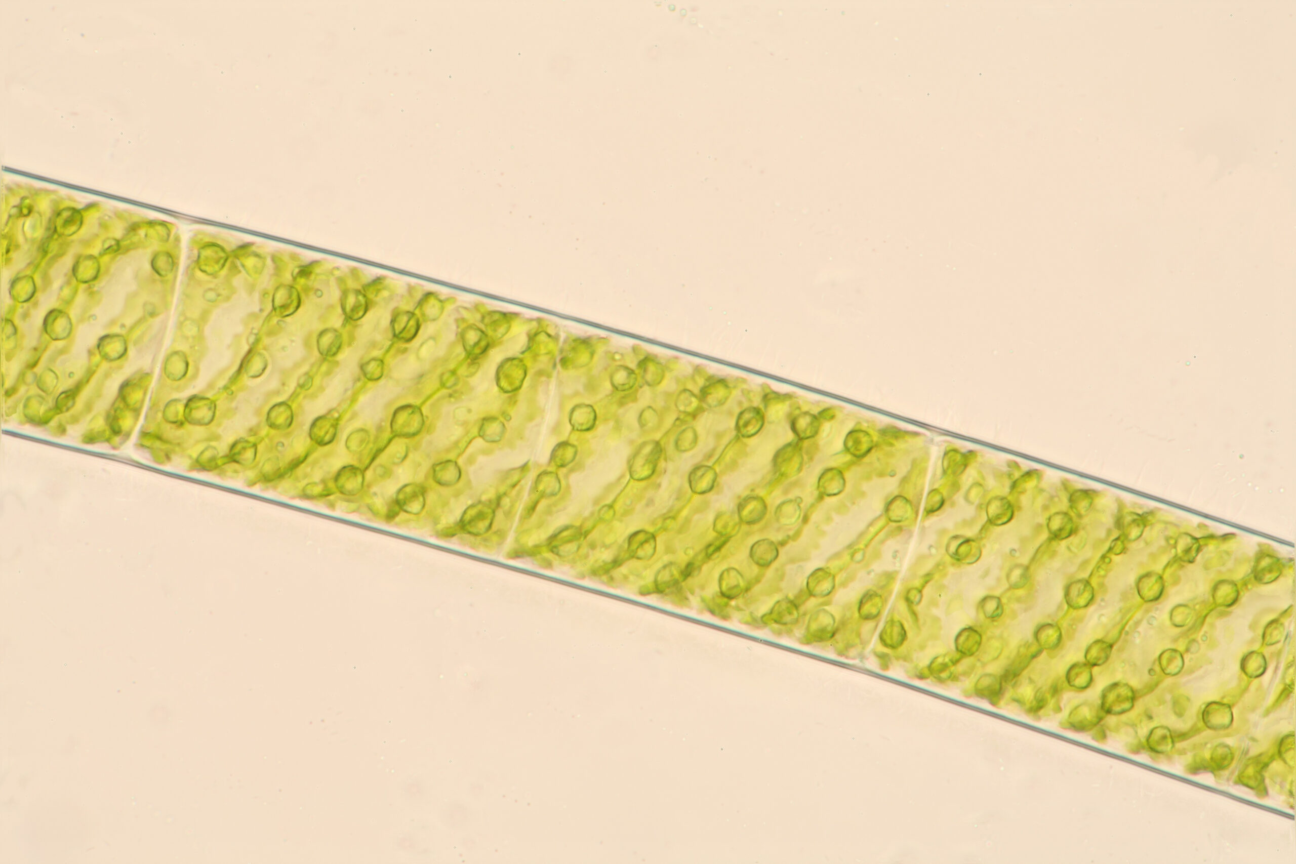
Single strand of Spirogyra
Seen from afar, algae is green slime. But up close it’s architecture.
This is a single strand of filamentous algae from the genus Spirogyra, characterized by its ribbon-like spiraling arrangement of chloroplasts.
Technical details
Olympus BH2, SPlan 40x 0.70, 2.5x NFK Photo eyepiece, Canon 5D Mark II. Focus stack of 55 images using Picolay. Sample from algae mat in stream, Bois de Boulogne, France.
Dancing diatoms
Diatoms are a mysterious group of unicellular creatures that look more like machines than living organisms. They build a glass shell around their bodies and glide around without any apparent form of locomotion.
Here a group of diatoms slide to and fro in a forest of algae. Presented here in a timelapse using polarized light.
Technical details
Olympus BH2, SPlan 10x 0.30, 2.5x NFK photo eyepiece, Canon EOS 5D Mark II. Cut out sheets of polarizing film used in trinocular head and above lamp. Pond water sample from park in Paris 16eme.
Algae floating with the current
Water doesn’t tend to move much on a microscope slide, but sometimes an air bubble will burst and everything will rush past in a tiny torrent. This single celled algae, part of the genus Mesotaenium characterized by its gelatinous capsule and leaf-shaped chloroplast, just went along for the ride.
Technical details
Olympus BH2, SPlan 100x 1.25 oil, 2.5x NFK photo eyepiece, Canon 5D Mark II. Rehydrated moss sample from Forêt de Fontainebleau, France.
Chasing a shapeshifter
I had a hard time keeping up with this little euglenid as it twisted and turned, showing off its ability to shapeshift, or exhibit what’s called metaboly. You can see clearly the flagella that it is using to propel itself along. I think this is a species of Peranema that has ingested some algae, but a confident identification has eluded me.
Also visible strands of filimentous algae and beaded strings of cyanobacteria
Technical details
Olympus BH2, SPlan 100x 1.25 oil, 2.5x NFK photo eyepiece, Canon EOS 5D Mark II. Pond water sample from park, Paris 16eme.
One creature’s waste…
This is a timelapse of an Amoeba Proteus performing exocytosis (or rather, defecating). You can see the material it leaves behind on the left of the frame, before it starts to make its way off to the right. Keeping a jar on my windowsill and taking samples from time to time reminds me that there is no such thing as waste in the microscopic world. Everything is used and reused in perpetuity.
Technical details
Olympus BH2, SPlan 40x 0.70, Olympus DCD 0.8-0.92, 2.5x NFK photo eyepiece, Canon EOS 5D Mark II. Sample from park pond in Paris 16eme.
Delicate engineering of the Thuricola valve
Thuricola is a genus of loricate peritrich, meaning a ciliate that anchors itself to feed and builds a protective shell around its soft body parts. The defining characteristic of this genus is the small “valve” or trapdoor that the individual zooids push out of the way when exiting the lorica. This valve, called an operculum, snaps shut when the zooids retreat, giving them protection against predators or debris.
In the second part of the video, we zoom in for a closer look at the moment the pair of zooids push past the operculum. Eventually this home gets too big for the two of them, and one undergoes a metamorphosis into a freely swimming organism and heads off to find a new place to settle down and build a home all over again.
Technical details
Olympus BH2, SPlan 40x 0.7 and SPlan 100x 1.25 oil, 2.5x NFK photo eyepiece. Canon 5D Mark II. Pond sample from park in Paris 16eme.
Anuraeopsis under pressure
Sometimes after searching a slide for little creatures for some time, the water starts to dry up so much that it traps organisms physically between the slide and the cover slip. A small drop of water releases the pressure, but before that it’s an opportunity to take a close look at organisms that are usually too fast to capture.
Here we see a single Anuraeopsis fissa, a species of rotifer that’s commonly seen carrying its own egg. Here we can clearly see the egg and how it’s tethered to the rotifer’s lorica. Also visible is a dinoflagellate from the genus Gymnodinium with its hard, walnut-shaped armored shell.
A drop of water added and the rotifer is freely swimming once again.
Technical details
Olympus BH2, SPlan 40x 0.70, 2.5x NFK photo eyepiece, Canon EOS 5D Mark II. Pond water sample from park in Paris 16eme.
Holophrya glides along while the tardigrade scrambles
Locomotion in the microscopic world takes many forms. Here we have a large single-celled ciliate from the genus Holophrya, characterized by its ovoid shape and large mouth at the tip of the organism. Ciliates glide along by beating rows of many small hairs called cilia.
We also see here a tardigrade, a multicellular creature with proper organs and a digestive system. Tardigrades move by crawling or scrambling along using their four pairs of clawed legs.
Technical details
Olympus BH2, SPlan 40x 0.70, 2.5x NFK photo eyepiece, Canon EOS 5D Mark II. Pond sample from park in Paris 16eme.
Even a flatworm can be cute
Cute isn’t the first word that comes to mind when we think of flatworms. But this Macrostomum gets points for having a proper set of eyes and a nose that it sticks into everything.
When we get closer, we see its body is covered in small spikes called rhabdite rods, mostly used for defense (distinct from its cilia which are for locomotion). The pharynx is also clearly visible, munching away. At the end we see the worm twisting and turning in polarized light, which enables us to see its muscle fibers as they stretch and contract.
Technical details
Olympus BH2, 10x SPlan 0.30 & 40x SPlan 0.70, 2.5x NFK photo eyepiece. Darkfield is with Olympus DCD 0.8 - 0.5. Polarized is with simple polarized film inserted in trinocular and above lamp. Canon EOS 5D Mark II. Pond water sample from park in Paris 16eme.